
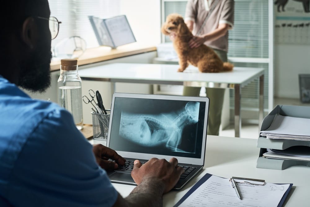
A dog x ray shows broken bones, swallowed objects, hidden dental disease, tumors, and organ problems that a physical exam alone cannot detect. This single imaging tool gives veterinarians a detailed look at what is happening beneath your pet’s skin, often within just a few minutes.
X-rays work by passing a short burst of radiation through the body. Dense structures like bone absorb that radiation and appear white on the image. Soft tissues let the rays pass through and show up in shades of gray, creating a clear map of your pet’s internal structures.
This article breaks down the four main categories of problems visible on veterinary radiographs: skeletal injuries, foreign body blockages, dental disease, and how modern digital imaging has changed the game for pet care.
Key Takeaways
- X-rays reveal bone fractures, arthritis, and skeletal tumors that guide surgical decisions.
- Radiography identifies swallowed objects and intestinal blockages requiring emergency care.
- Dental radiographs expose hidden tooth root damage and jawbone disease invisible during visual exams.
- Modern digital systems deliver instant results with minimal radiation exposure.
- Common concerns about safety, sedation, and cost answered below.
Can a simple x-ray really catch a broken bone your vet missed by touch?
X-rays transform invisible injuries into clear images within minutes. When dogs experience falls, car accidents, or rough play, radiographs pinpoint the exact fracture location and severity. Veterinary radiography remains the most frequently used form of medical imaging in clinical practice, as described by the UC Davis Veterinary Medical Teaching Hospital.
Hairline fractures are sneaky. They cause subtle limping that many owners dismiss as soreness or a pulled muscle. However, without imaging, these small cracks can worsen into full breaks that require much more invasive treatment.
Radiographs catch these hairline injuries before they progress. The images reveal exactly where the bone has cracked, how the fragments line up, and whether nearby soft tissue took damage too.
Complex fractures with multiple bone fragments look like scattered puzzle pieces on a radiograph. Surgeons rely on these detailed images to plan their approach. They use the X-ray as a roadmap for placing pins, plates, and screws in the right positions for stable healing.
Joint Conditions Hiding in Plain Sight
Hip dysplasia appears on film as shallow hip sockets that fail to hold the femoral head securely. This instability leads to cartilage wear and painful bone changes over time.
Cruciate ligament tears shift how the tibia sits relative to the femur. Radiography remains one of the most commonly used diagnostic tools for identifying these joint abnormalities and choosing between surgical repair or conservative care, as outlined in the Merck Veterinary Manual.
Bone Tumors and Arthritis on Film
Radiographs also pick up signs of bone cancer and chronic joint disease:
- Osteosarcoma appears as aggressive bone destruction mixed with abnormal new growth patterns
- Early detection through imaging improves treatment outcomes and may slow cancer spread
- Arthritis shows as joint space narrowing, bone spurs, and cartilage loss over time
- Sequential X-rays help veterinarians track disease progression and adjust pain management plans
Your dog swallowed what? How X-Rays Expose Dangerous Blockages
Dogs explore the world with their mouths, and swallowing the wrong item can turn life-threatening fast. Gastrointestinal foreign body obstructions are emergencies often requiring surgical removal, as reported by the Cornell Riney Canine Health Center. Radiographs reveal exactly what your dog ate and where the object sits in the digestive tract.
Not every material shows up the same way on film. Here is what veterinarians typically see:
- Metal objects, coins, and jewelry glow bright white on the image
- Cooked bones appear white but may fragment into sharp, gut-piercing pieces
- Dense rubber and plastic toys create visible gray shadows
- Rocks and hard materials stand out clearly against soft tissue backgrounds
When a foreign object lodges in a narrow intestinal passage, digestion stops and gas builds up behind the blockage. These trapped gas pockets appear as dark areas on the image, forming a recognizable pattern that signals an immediate surgical emergency.
Some materials are trickier to spot. String, fabric, and certain soft plastics absorb radiation similarly to the body’s own tissues. In these cases, veterinarians use contrast studies. Your pet swallows a barium or iodine-based liquid that coats the digestive tract lining and reveals obstructions standard images miss.
Bloat, Stones, and Heart Problems on X-Ray
Gastric dilatation and volvulus, commonly called bloat, ranks among the most dangerous emergencies for large breed dogs. This condition occurs when the stomach fills with gas and rotates on the mesenteric axis, cutting off blood supply, as detailed in the Merck Veterinary Manual. X-rays confirm the diagnosis by showing a distinctive double-bubble pattern in the stomach.
Bladder stones and kidney stones show up as bright white objects in the urinary system. The images reveal stone size, number, and exact location. Heart conditions also become visible as enlarged cardiac silhouettes on chest radiographs, pointing to problems that need prompt treatment.
80% of Dogs Have This Dental Problem, and You Can’t See It Without X-Rays
Most dental disease hides below the gum line where a standard visual exam cannot reach. Dental radiography is a core diagnostic tool for detecting hidden, painful pathology that would otherwise go untreated for months or even years, as published in the Journal of Veterinary Dentistry.
Periodontal disease affects a large majority of dogs by age three, yet the worst damage stays completely invisible on the surface. Dental radiographs expose bone loss around tooth roots that indicates advancing infection. These images show exactly how much supporting bone remains, helping veterinarians decide which teeth still have a chance and which ones need to come out.
Fractured Roots and Tooth Resorption
Tooth resorption eats away at tooth structure from the inside, causing severe pain your dog cannot communicate. Dental radiographs reveal these hidden cavities as dark spots inside normally solid teeth, often catching the condition long before any external sign appears.
Fractured roots from chewing hard objects or blunt trauma may look perfectly normal during a visual exam. X-rays tell the real story. They show dark lines running through the tooth and reveal whether the fracture extends into the nerve.
Jawbone Growths and Hidden Teeth
Tumors and cysts can develop silently in the jawbone supporting your pet’s teeth. Dental radiographs help veterinarians separate routine gum problems from serious growths that require immediate surgical attention.
Un-erupted teeth trapped inside the jawbone can form cysts that destroy surrounding bone over time. Even more concerning, chronic oral infections may spread through the bloodstream to the heart, kidneys, and liver. This connection between mouth health and whole-body health makes regular dental imaging a critical part of preventive veterinary care.
Digital X-Rays Changed Everything, Here’s Why Your Vet Switched
Modern veterinary clinics now use digital radiography systems that capture images in seconds and display them instantly on a screen. This technology does not require anesthesia for most patients. It eliminates the chemical processing delays of old-fashioned film X-rays, as highlighted by the UC Davis School of Veterinary Medicine.
Here are the key advantages digital systems bring to your pet’s care:
- Reduced radiation exposure makes repeat imaging safer for monitoring chronic conditions
- Images can be brightened, zoomed, and enhanced without retaking the X-ray
- Digital files store permanently for comparing current results against future exams
- Veterinarians can share images with specialists worldwide in minutes
Senior dogs with arthritis benefit greatly from this technology. Their veterinarians can monitor joint changes regularly without worrying about cumulative radiation exposure. Digital archives let them overlay old and new images side by side, catching subtle shifts in bone density or organ size over time.
When standard radiographs do not provide enough detail, advanced modalities step in. CT scans create cross-sectional images that show internal structures in fine detail. MRI offers excellent soft tissue contrast for brain, spinal cord, and joint problems. Contrast studies highlight blood vessels, kidneys, and the digestive tract to reveal issues that routine X-rays simply cannot capture.
Why Every Vet Reaches for Digital X-Rays First
Dog x ray technology gives veterinarians a fast, detailed view inside your pet’s body that no physical exam can match. From hidden fractures and swallowed toys to silent dental disease and early-stage tumors, radiographic imaging catches problems when treatment is most effective and least invasive.
Digital systems now deliver instant, high-resolution results with lower radiation than ever before. Routine imaging during wellness visits can spot trouble long before your pet shows obvious symptoms, saving both stress and money in the long run.
If you have questions about what imaging can reveal for your pet, learn more about our digital radiology and X-ray services and reach out to our team to discuss your options and schedule an appointment.
FAQs
How much radiation exposure do dogs receive during X-rays?
Modern digital systems use very low radiation doses that pose negligible health risks. The amount your dog receives during a typical exam compares to just a few days of natural background radiation. Veterinarians also use protective shielding and limit the number of images to keep exposure as low as possible.
Can X-rays detect soft tissue problems like tumors in organs?
X-rays can reveal some soft tissue abnormalities, like fluid in the lungs or an enlarged heart. However, CT scans and ultrasound provide much better visualization of organ tumors and internal soft tissue problems. Radiographs work best for bones, dense foreign objects, and certain chest or abdominal conditions.
Will my dog need anesthesia for X-ray procedures?
Most calm, cooperative dogs tolerate positioning without any sedation. Anxious dogs or those in significant pain may need mild sedation so the veterinary team can get clear, properly positioned images. The decision depends on your pet’s temperament, the body area being examined, and how many views are needed.
How quickly can veterinarians get X-ray results?
Digital systems display images on screen within seconds of taking them. Your veterinarian can review and interpret results right away, often while you wait. This rapid turnaround allows immediate treatment decisions during emergencies and cuts waiting time for routine diagnostic appointments.
What should I expect to pay for dog X-rays?
Costs vary depending on the number of images, body area, and whether it is a routine or emergency visit. Emergency imaging typically runs higher than a planned diagnostic session. Dental radiographs and specialized contrast studies may carry separate fees, so ask your veterinary team for an estimate before the appointment.
Sources:
- https://www.vetmed.ucdavis.edu/hospital/diagnostic-imaging/small-animal/xray
- https://www.merckvetmanual.com/clinical-pathology-and-procedures/diagnostic-imaging/overview-of-diagnostic-imaging-of-animals
- https://www.vet.cornell.edu/departments-centers-and-institutes/riney-canine-health-center/canine-health-topics/gastrointestinal-foreign-body-obstruction-dogs
- https://www.merckvetmanual.com/digestive-system/surgical-problems-of-the-gastrointestinal-tract-in-small-animals/gastric-dilation-and-volvulus-in-small-animals
- https://pubmed.ncbi.nlm.nih.gov/19410234/
- https://ceh.vetmed.ucdavis.edu/imaging
